The low frequency ESR apparatus is useful for measuring spin susceptivility of organic substances, especially with Pauli paramagnetism. As you know, SQUID susceptometer is superier in sensitivity, but it cannot descriminate Pauli paramagnetism from the diamagnetic contributions of the substance, both of which are usually temperature independent. However, ESR observes only spin magnetisms, but not diamagnetisms, which is a clear advantage over the SQUID susceptometer.
One usually uses an electromagnet with iron cores: ESR is measured at low magnetic field and then, its intensity is calibrated with 1H NMR signal intensity measured simply by increasing the magnetic field, which satisfies the NMR resonance condition. We call these procedures as the ESR-NMR technique. This is a good way, but it is a relatively time consuming procedure, There are mainly two reasons:
- To keep accurate, the amplitude of the field modulation to observe a derivative signal of the absorption spectrum should be sufficiently lower than the spectral line width, resulting in the suppression of sensitivity in ESR and NMR measurements. As a result, lots of accumulations of signals are required for us to achieve an enough S/N ratio.
- There is a significant limitation in the measurement conditions: Magnetic field scans to accumulate signals should be enough slow to keep the scan rate unchanged because of the iron hysteresis of the electromagnet with iron cores.
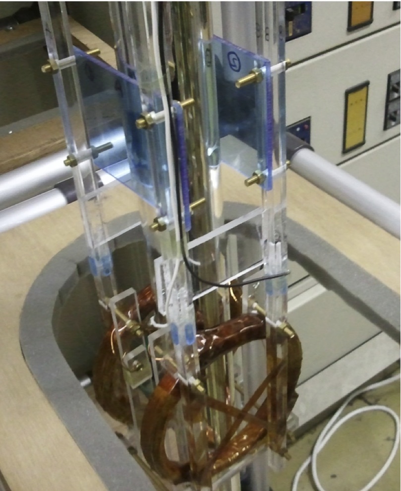
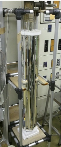
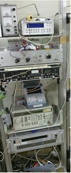
The low frequency ESR system shown in the above photographs has several advantages over the usual ESR-NMR technique. Characteristics are summarized as follows.
- Measures only ESR as functions of temperature and pressure, and calibrate by comparing it with that of a standard sample, for example, DPPH.
- Magnetic field is produced by an empty electromagnetic coil without any cores, which is basically independent of the hysteretic distortions in ESR baselines, even in fairly rapid scan rates of the magnetic field.
- ESR absorption spectra are measured without the magnetic field modulation.
The rapid scan rates make us possible to measure the absorption spectra without the field modulation, which is a requirement to suppress the 1/f noise. We can use the scan rate higher than 10 Hz without any distortion of the signals. Since both of the sample and the standard sample are measured in a sample tube simultaneously, the linewidth of the standard sample should be sufficiently differrent from that of the sample. If the number of spins in the samples are the same as each other, the intensity ratio of the ESR absorption signals is proportional to the ratio of the linewidths, which is markedly different from the square of the linewidth ratio in the derivative signals with the magnetic field modulation.
As a result of the above factors, the consumption time of the measurements with the present system is much less than that with the ESR-NMR technique, 1/10-1/100 or more.
- Intensity of the absorption in the sharp signal is more than three times larger than that of the derivative signal with the modulation, since the amplitude of the modulation should be less than 1/3 of the linewidth. Intensity of the broad signal measured with the same modulation amplitude as the sharp one is further weak by the ratio of the linewidth, which corresponds to more than 1/10 in total.
- Scan duration for the ESR-NMR technique is around 60 s for up scan, which is 600×2 times longer than the present system with a 10 Hz scan for up and down scans. Thus, the final ratio of the S/N ratios reaches square root of 1,200, that is ~1/35.
Thus, this system saves the measurement time by 1/10×1/35. The magnetic field coil is located in a liquid nitrogen space to reduce the Joule heating, which is driven by a 60V-2.5A constant current power supply in the lowest part of the rightmost picutre. ESR frequency is 150-200 MHz with the magnetic field of 5-7 mT.
Development of a similar system for the cubic amvil cell up to 8 GPa is under way.